
Long distances between reference points
Between implants, there may be sections with few unambiguous geometric features. The scanner must stitch the partial record across a greater distance.
Technical guide · digital full-arch scanning
One clinical pathway connects the Multi-Unit abutment (MUA) platform, ScanLogiQ, AssistLogiQ, Scan Transfer and clinical passive-fit verification. This is not a component catalogue, but an overview of decision and control steps in digital full-arch scanning.
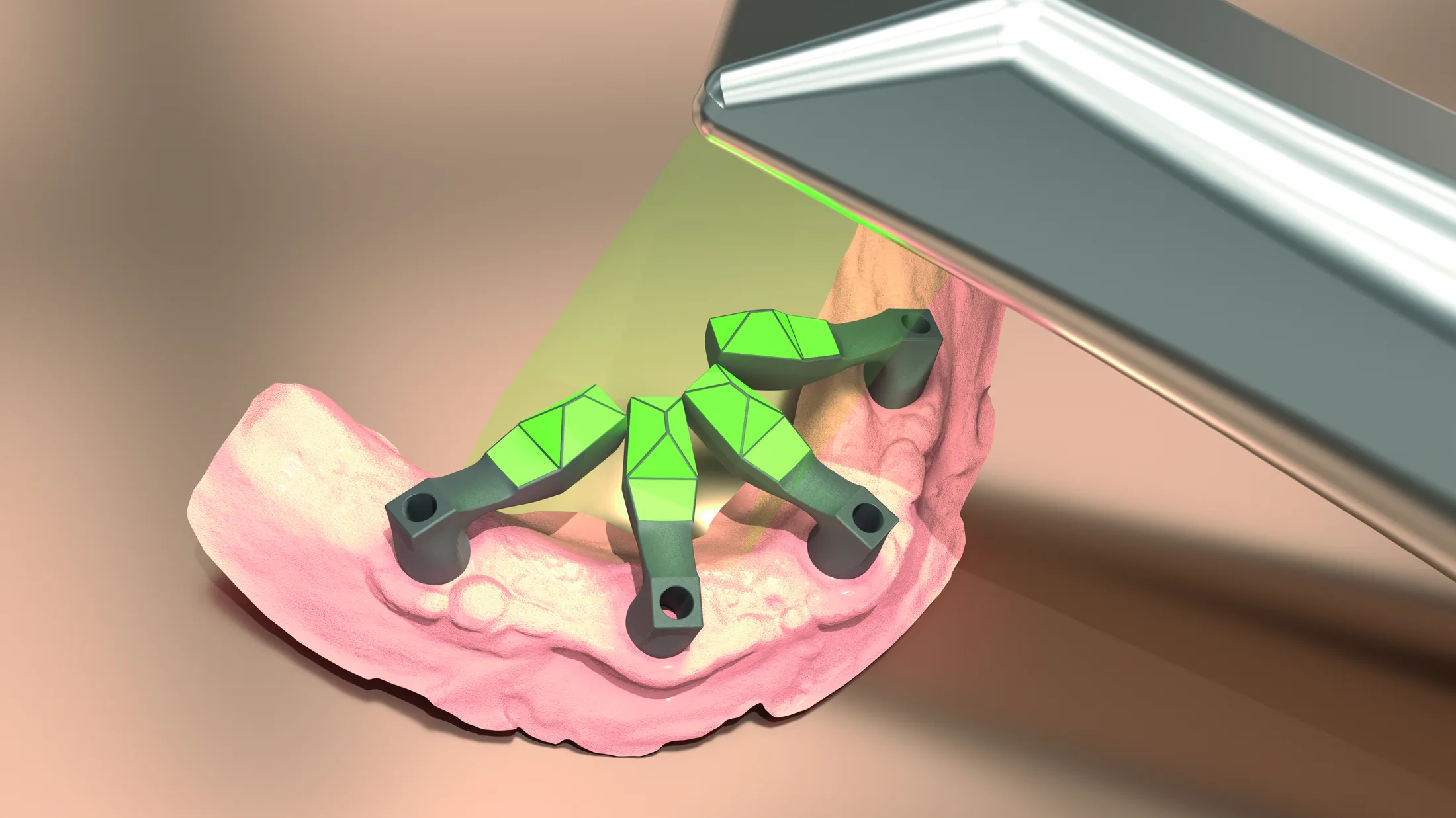
Stitching drift, four independent Scan Flag iQ acquisitions, Data Sets 1–3, AssistLogiQ, 6DoF and the limits of digital validation. Matouš Záhorec, a professional trainer with extensive experience in CAD/CAM technologies and implant prosthodontics, will add a clinical-laboratory perspective to the live discussion.
Why discrepancies occur
In an extensive implant-supported restoration, the intraoral scanner must connect multiple distant reference points into one spatial whole. The longer the scan path and the fewer stable references available to the software, the more important it becomes to verify the data repeatedly.
Between implants, there may be sections with few unambiguous geometric features. The scanner must stitch the partial record across a greater distance.
Soft tissues, saliva, reflections and pronounced curvature make stable acquisition more difficult, especially in distal areas and locations with poorer visibility.
Small local inaccuracies can accumulate as successive captures are stitched together. The resulting model may still look smooth and convincing on screen.
Trueness describes how close the record is to the true position. Repeatability (precision) describes whether repeated scans produce a similar result. ScanLogiQ works with repeated acquisitions precisely so that data that look good but are not mutually consistent can be identified.
Guide map
Each phase answers a different question. If a control point is missing, a discrepancy can pass through to the definitive restoration. The workflow is therefore divided into clear verification checkpoints.
Platform
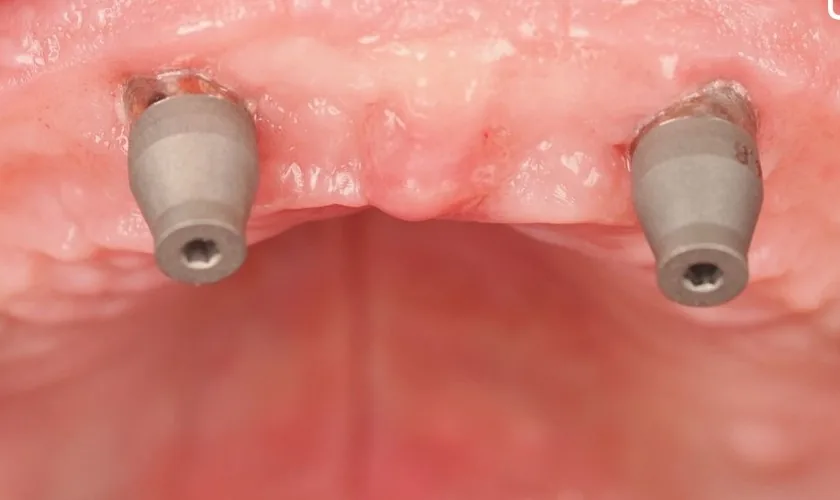
Implant-level connections differ between systems. A Multi-Unit abutment (MUA) creates a common prosthetic level above the implants, allowing scanning, framework design and communication with the laboratory to be standardised.
MUA AP availability must be verified for the specific implant platform. The general principle of working at MUA level should therefore be distinguished from the availability of individual AP references.
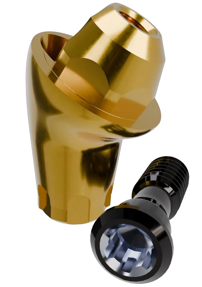 MUA AP · anatomically shaped external profile
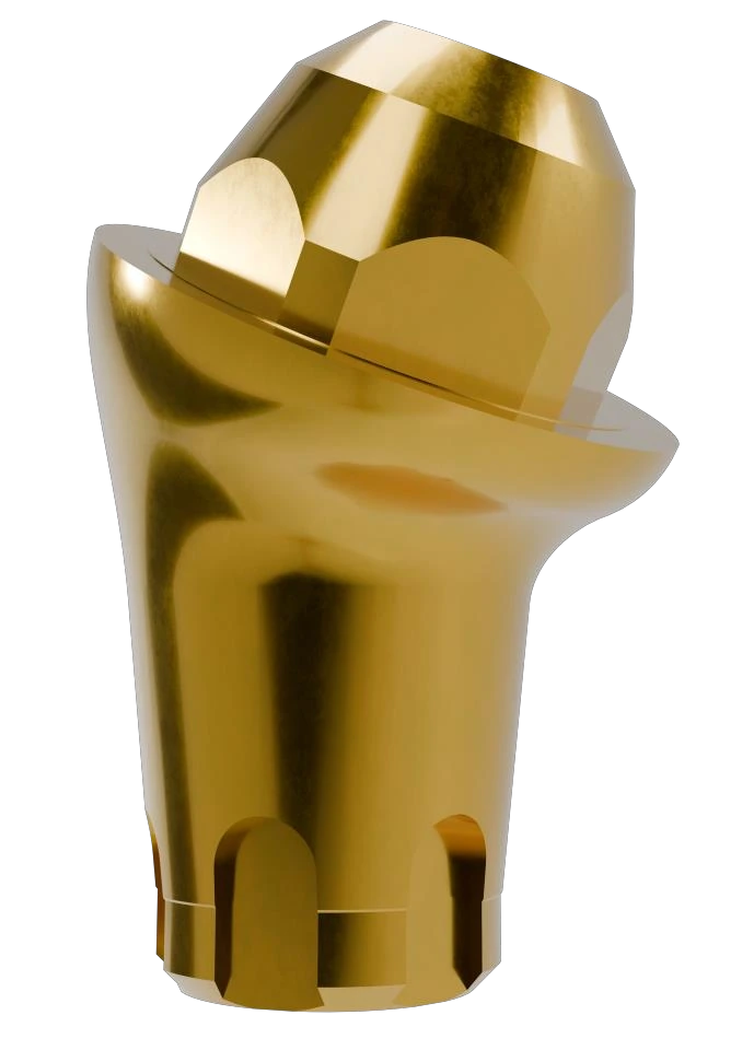 overall shape
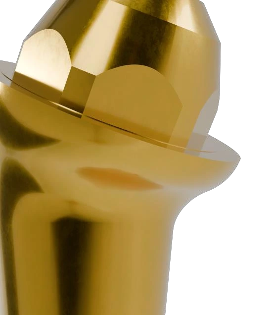 profile detail
The curved MUA AP profile is designed to provide a smoother transition between the abutment and soft tissues.
The gold-coloured titanium nitride (TiN) surface combines an aesthetic appearance with high surface wear resistance.
Angulated versions allow correction of an unfavourable implant axis and can improve access to the restoration screw channel.
ScanLogiQ
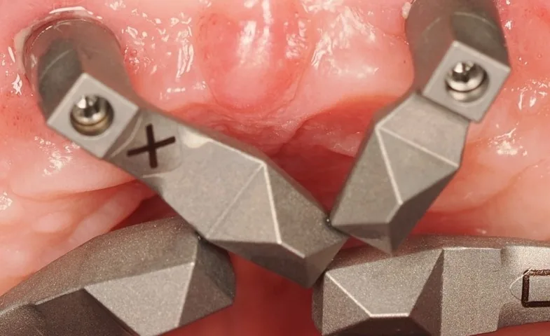
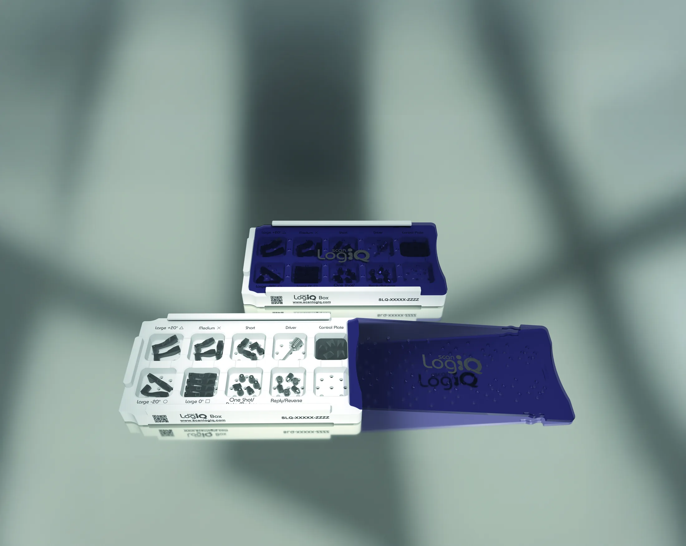
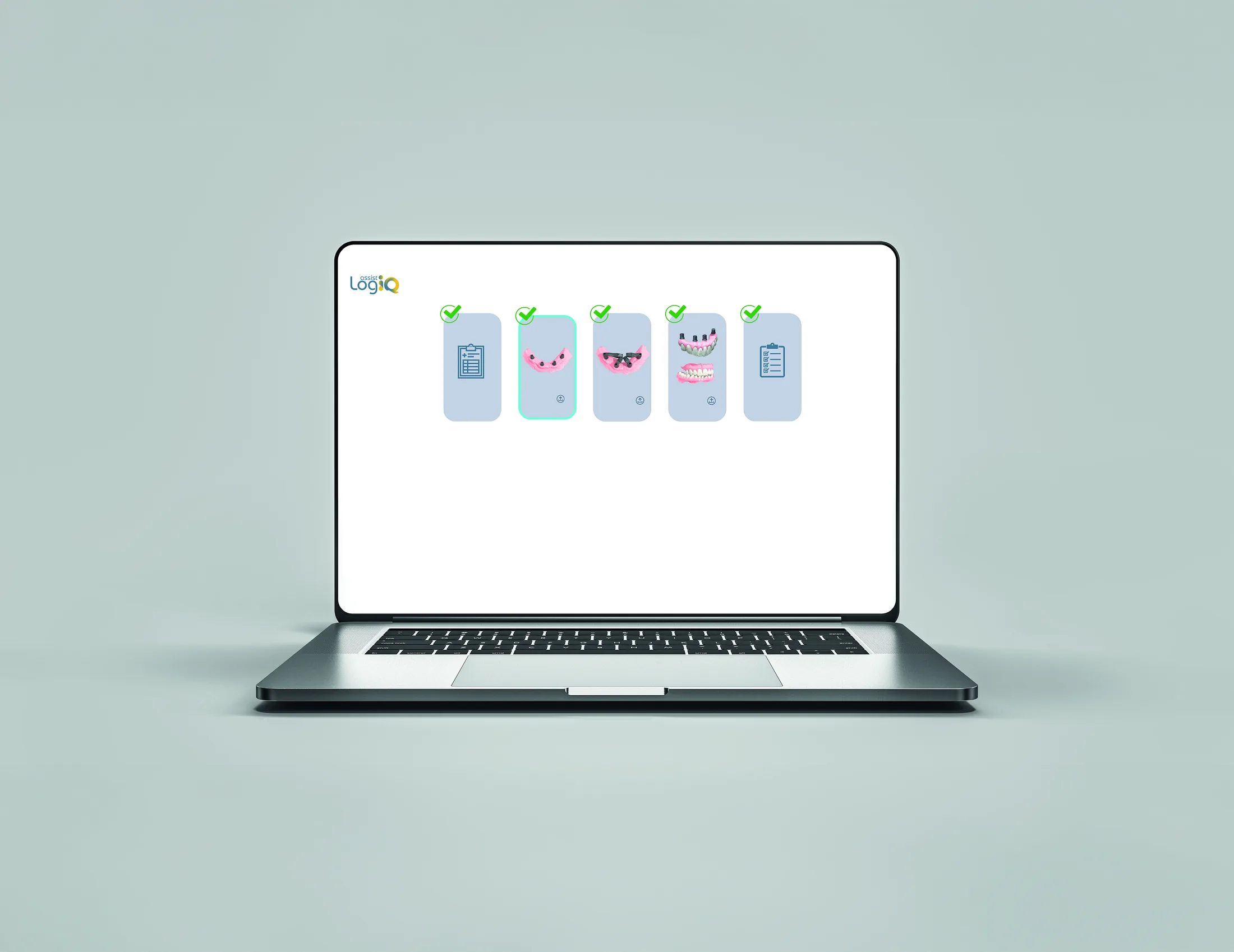
ScanLogiQ combines the physical Scan Flag iQ components with the AssistLogiQ platform. The aim is to record the MUA position repeatedly and identify inconsistent data before they are used to design the definitive restoration.
Captures the soft tissues and the initial map used to select and orient Scan Flag iQ.
Four independent acquisitions make it possible to assess whether the spatial information repeats consistently.
The number of mutually confirmed scans determines whether it is appropriate to proceed or repeat the acquisition.
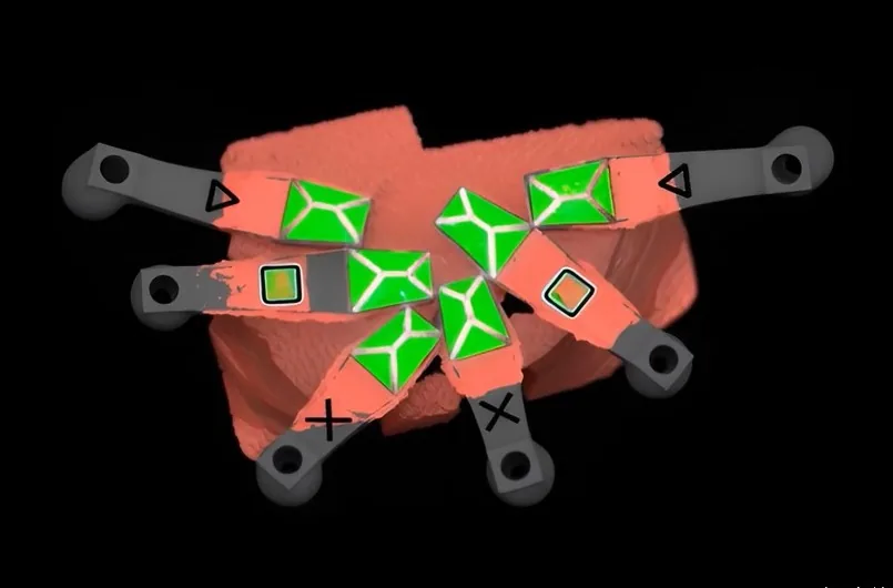
AssistLogiQ evaluation
Repeating a scan when consistency is low is not a failure; it is part of quality control. Four confirmed files represent a complete ScanLogiQ validation result, although the passive fit of the definitive restoration must still be verified clinically.
| Result | Meaning of the result | Recommended action |
|---|---|---|
| 1–2 / 4 | Insufficient consistency | Repeat the scan; depending on case risk, consider a physical verification framework. |
| 3 / 4 | Minimum acceptable consistency | Add another scan or proceed according to the clinical risk and internal protocol. |
| 4 / 4 | Complete consistency across repeated acquisitions | Proceed to design and manufacture while retaining subsequent clinical passive-fit verification. |
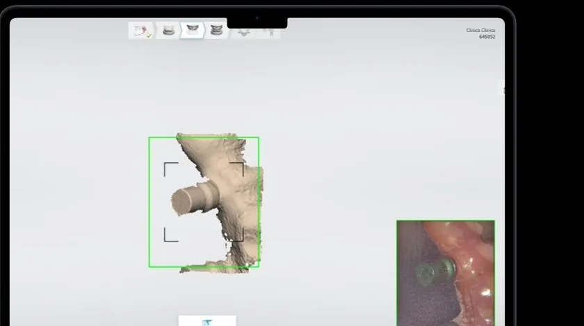
Reverse iQ
Scan Flag iQ is used to verify spatial position at MUA level. Reverse iQ can add the shape, functional relationships, occlusion and soft-tissue support from a provisional restoration, prototype or try-in. It is therefore an optional but valuable data layer in many cases.
Scan Transfer
Scan Transfer uses a physical reference structure and allows rigid splinting of the scan bodies. ScanLogiQ is based on repeated controlled scanning and software evaluation. Both approaches target the same problem: limiting the accumulation of deviations during intraoral scanning of a long dental arch.
Connected scan bodies create a continuous reference path along which the intraoral scanner can move without large jumps between isolated points.
The rigid assembly can serve as a physical validation key to verify position and mutual relationships before definitive manufacturing.
The simpler portion is recorded intraorally, while a difficult-to-access area can be rescanned extraorally and then accurately aligned.
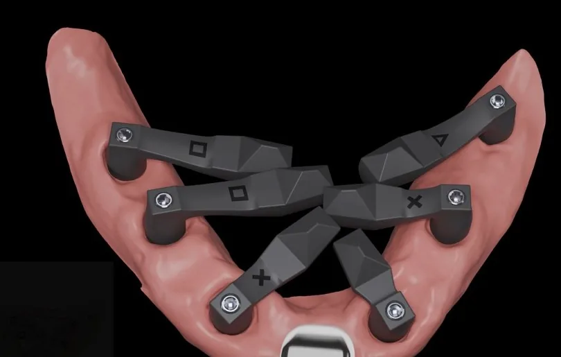
Clinical verification
After data evaluation and manufacture of the restoration, clinical verification follows. The Sheffield test and radiographic verification complement the digital workflow and confirm whether the restoration actually seats passively in the mouth.
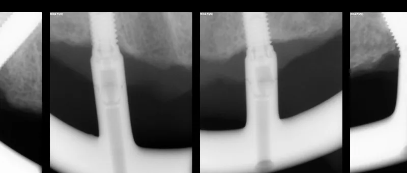
Scientific resources
For each technical claim, it is useful to state what the study actually tested, which reference method it used, what results it found, and which conclusions cannot be generalised from it.
Revilla-León et al. evaluate an uncalibrated system of scan bodies oriented towards the centre of the arch using five intraoral scanners. DOI: 10.1016/j.prosdent.2026.03.002.
A published clinical protocol describes continuous scanning and rigidly connected scan bodies as a reference pathway for digital full-arch recording.
The clinical description validating the principle presents the CSS, De|Bug and Re|Scan procedures. It is suitable for explaining the method, not as universal evidence for every clinical situation.
Study results must be interpreted in relation to the specific procedure tested, the reference method and the measurement conditions. Indirect rankings of different technologies cannot be presented as direct comparisons.
Check before first clinical use
Before the first clinical use, it is advisable to run through the entire workflow: intraoral-scanner setup, STL file saving, Data Set 1, four Data Set 2 scans, evaluation, optional Reverse iQ, Data Set 4 and download of the resulting working files.
27. 11. 2026 · Žďár nad Sázavou
At the Digitally to the Perfect Result conference, ScanLogiQ will be explored from trueness vs. precision through stitching drift, four Scan Flag iQ acquisitions and AssistLogiQ to the limits of digital validation. The planned schedule also includes an interactive “Kill the workflow” conclusion and an optional hands-on block.
Related technical content